Home
/ Labeled Diagram Of An Eye, Vision And Eye Diagram How We See : Introductionintroduction the eye is a specialized sense organ that helps us to understand our environment.
Labeled Diagram Of An Eye, Vision And Eye Diagram How We See : Introductionintroduction the eye is a specialized sense organ that helps us to understand our environment.
Labeled Diagram Of An Eye, Vision And Eye Diagram How We See : Introductionintroduction the eye is a specialized sense organ that helps us to understand our environment.. Contrary to popular belief, the eyes are not perfectly spherical; Lungs and respiratory system for kids gcse science breathing and. For us to see, there has to be light. Basic eye anatomy understanding the anatomy of the eye is critical to understanding cataracts, how and why cataracts affect vision, and cataract surgery itself. Handout illustrating parts of the eye keywords:
It is the transparent membrane which refracts the light entering our eye. The human eye is composed of many different parts that work together to interpret the world around us. Select one anterior chamber ciliary body cornea fibrous tunic iris lateral rectus muscle lens medial rectus muscle optic disk optic nerve pupil retina vascular tunic vitreous nerve. Instead, it is made up of two separate segments fused together. An eye diagram triggered from a clock recovered from the data signal using a narrow loop bandwidth clock recovery scheme.
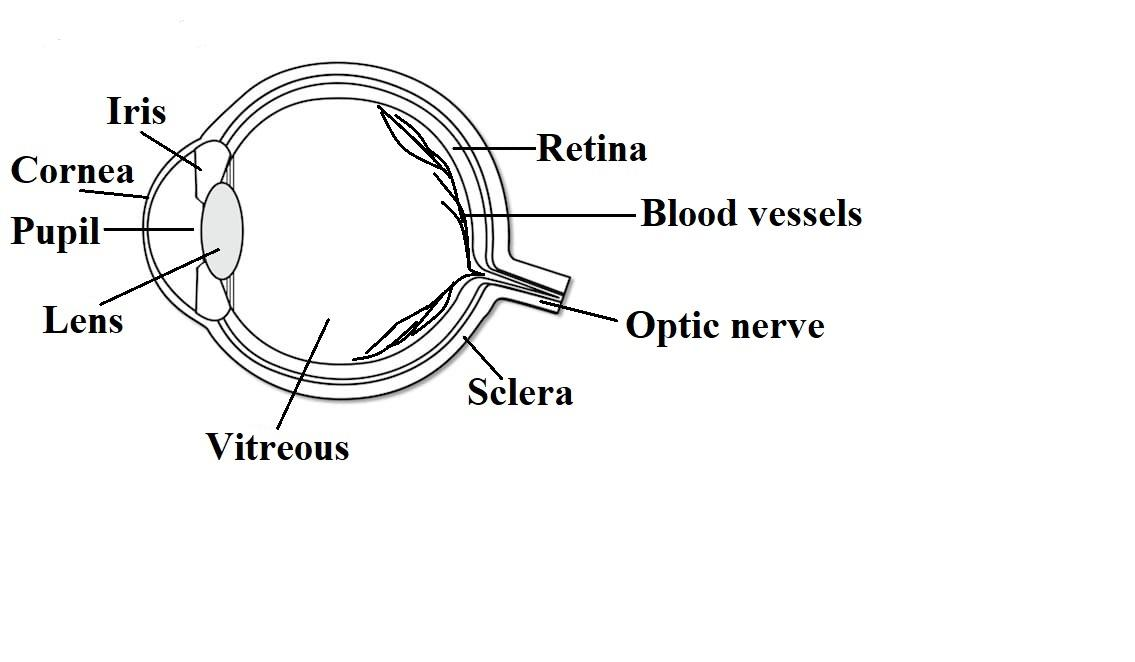
Parts Of The Eye Their Function Robertson Optical And Optometry from www.md-health.com Diagram of human eye with labelling. Labeled diagram of the eye The anterior part of this layer is called cornea. It is the outer covering, a protective tough white layer called the sclera (white part of the eye). When light shines on an object, a reflection is sent which passes through the eye lens and later projects the image of the. This is a small tube that runs from the eye to the nasal cavity. Introductionintroduction the eye is a specialized sense organ that helps us to understand our environment. At the front of the eye is a clear, round window called the cornea.
The core eye anatomy diagram, designed as the labeling exercise, has a fully colored and labeled reference chart to go with it.
It is the outer covering, a protective tough white layer called the sclera (white part of the eye). Lungs and respiratory system for kids gcse science breathing and. Select one anterior chamber ciliary body cornea fibrous tunic iris lateral rectus muscle lens medial rectus muscle optic disk optic nerve pupil retina vascular tunic vitreous nerve. Let's take a closer look at each of these. Contrary to popular belief, the eyes are not perfectly spherical; Tear drains from the eyes in to the nose through the tear duct. Human eye anatomy quiz diagram labeling, eye anatomy model, interactive eye diagram quiz. Iris, optic nerve, pupil, cornea, lens, retina. Simple anatomy of the retina by helga kolb. Although the eye is small relative to most organs in the human body, it has many distinct anatomical parts, all of which contribute to the production of normal vision in one way or another. This helps us to understand how each one is situated and related to the other. The anterior part of this layer is called cornea. With help from other important structures in the eye, like the iris and cornea, the appropriate amount of light is.
Diagram of human eye with labelling. Read the definitions, then label the eye anatomy diagram below. It consists of the following parts: Draw a well labelled diagram of eye. Receptor, sensory pathway, and a brain center.
Draw A Labelled Sketch Of The Human Eye Class 12 Physics Cbse from www.vedantu.com Introductionintroduction the eye is a specialized sense organ that helps us to understand our environment. Handout illustrating parts of the eye keywords: The anterior part of this layer is called cornea. One of our favorite ways to get to grips with all of the parts of the eye is by utilizing labeled diagrams. This is why a teary eye is usually accompanied by a runny nose. You should make a label that represents your brand and creativity, at the same time. Vision is by far the most used of the five senses and is one of the primary means that we use to gather information from our surroundings. At the front of the eye is a clear, round window called the cornea.
Fungi diagram gcse top electrical wiring diagram.
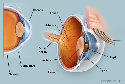
Six extraocular muscles in the orbit are attached to the eye. Causes loss of central vision as you get older. On a diagram of the eye, we can see all of the relevant structures together on one image. Eye anatomy, eye diagram, cornea, iris, lens, macula, optic nerve, pupil, retina, vitrous gel, diabetic eye disease. From the center of the optic nerve radiates. Human eye anatomy quiz diagram labeling, eye anatomy model, interactive eye diagram quiz. Basic eye anatomy understanding the anatomy of the eye is critical to understanding cataracts, how and why cataracts affect vision, and cataract surgery itself. Lungs and respiratory system for kids gcse science breathing and. Often called lazy eye, this condition starts in childhood.one eye sees better than the. The cornea, the pupil, the iris, the lens, the vitreous humor, the retina, and the sclera. It allows the light entering our eye to pass through it. When an ophthalmologist uses an ophthalmoscope to look into your eye he sees the following view of the retina (fig. A human eye is roughly 2.3 cm in diameter and is almost a spherical ball filled with some fluid.
Read the definitions, then label the eye anatomy diagram below. It is the outer covering, a protective tough white layer called the sclera (white part of the eye). An eye diagram triggered such that the delay between jittered clock and jittered data destructively interferes. Facts about the eye to understand more in detail about our eye and how our eye functions, we need to look into the structure of the human eye. Our latest youtube film is ready to run.
The Eyes Human Anatomy Diagram Optic Nerve Iris Cornea Pupil More from img.webmd.com It is a sensory unit composed of three parts: National eye institute , national eye health education program subject: What you want to interpret as a major part of the human eye is somewhat up to the individual, but in general there are seven parts of the human eye: Eye anatomy, eye diagram, cornea, iris, lens, macula, optic nerve, pupil, retina, vitrous gel, diabetic eye disease. Eye anatomy complete physiology of eye is described below in the given paragraph:. Vision is by far the most used of the five senses and is one of the primary means that we use to gather information from our surroundings. This is an exercise for students to label a simple blank eye diagram with the following parts: From the center of the optic nerve radiates.
On a diagram of the eye, we can see all of the relevant structures together on one image.
Label parts of the human eye. This is why a teary eye is usually accompanied by a runny nose. Lungs and respiratory system for kids gcse science breathing and. Instead, it is made up of two separate segments fused together. The anterior part of this layer is called cornea. Contrary to popular belief, the eyes are not perfectly spherical; Human eye consists of various parts which helps us in seeing the objects, the function of various parts are: The cornea, the pupil, the iris, the lens, the vitreous humor, the retina, and the sclera. This is an exercise for students to label a simple blank eye diagram with the following parts: Let's take a closer look at each of these. The core eye anatomy diagram, designed as the labeling exercise, has a fully colored and labeled reference chart to go with it. Handout illustrating parts of the eye keywords: Select one anterior chamber ciliary body cornea fibrous tunic iris lateral rectus muscle lens medial rectus muscle optic disk optic nerve pupil retina vascular tunic vitreous nerve.